Diatom of the Month: November 2016 - Medlinella amphoroidea
by Tom Frankovich*
I would like to introduce you all to Medlinella amphoroidea, a new taxon that was observed on loggerhead
sea turtles, as the November diatom-of-the month. But, before
I get to discussing the morphology and ecology of this new genus and species, I will tell you all a personal story of serendipity
and professional relationships. It was early 2013, and I had received an
email from Dr. Brian Stacy, a marine veterinarian at the National Marine Fisheries Service, and
a friend from when we worked together investigating parasites in marine
gastropods. Brian told me that his wife, Dr. Nicole Stacy, also a marine
veterinarian, was interested in identifying organisms that she suspected were
diatoms that were on skin smear slides and contaminants in blood, urine and
teat fluid samples from Florida
sea turtles and manatees.
Fig.
1. Dr. Brian Stacy performing a necropsy on a loggerhead turtle, Caretta caretta (Photo courtesy
Brian Stacy, unknown photographer).
I subsequently found out that pathologists were frequently
misidentifying diatoms and reporting them as “parasite eggs”! Obviously, it is
important to distinguish likely benign diatoms from harmful parasites, and so I
told Nicole that I would gladly examine photomicrographs of her samples and that
it would be no problem to identify these suspected diatoms using descriptions
of the local diatom flora. After all,
the sea turtle and manatees probably picked up these diatoms from the
surrounding environments, right? Wrong! Nicole had immediately sent me
images of various samples collected from manatees and sea turtles. The samples
were uncleaned and were stained with a dye to reveal cytological
characteristics of interest to a pathologist. These are not the best samples
for a diatomist to examine, so most diatoms could only be identified in the
broadest of terms (e.g., radial centric,
raphid pennate). I
asked Nicole if she had material to clean, mount, and examine using standard
diatom methodologies. She told me that she only receives prepared slides from
the field. End of story? Not yet. About
a week had passed when an Everglades Park Ranger knocked on my office door at
our Florida Bay research station and asked if I could help him move a dead
manatee that was reported in the bay. I suppose most people would say no to
moving a dead smelly manatee, but for me this was a gift from the heavens.
I finally got my hands on an adequate sample for my planned examinations, but I
was in for a rapid deflation of my presumed diatom identification abilities,
and for a big surprise.
Fig.
2. A manatee captured for a health assessment (left) and a close –up of the
manatee skin (right) with a film of epibionts, including diatoms (Photos by Tom Frankovich).
If you are a fellow diatomist in the blogosphere, you may
agree with me that the most exciting part of our work is looking at a sample
for the first time. Like a child waiting
for Christmas morning, I anxiously awaited for the cleaning and rinsing of the
new diatom sample to be complete. What I saw through the microscope was an
assemblage unlike anything I had seen previously. First, instead of seeing a
very diverse collection of tens of diatom taxa, I saw an assemblage comprised
of very few taxa. 95% of the valves
appeared to belong to 2 or 3 taxa. Second, I could not identify the dominant
taxa, not even to a genus! Even
after scouring 58 reference books, and a file cabinet of reprints of benthic
diatom taxonomy, I was still lost! Time to call for help. I emailed
photomicrographs to Dr. Mike Sullivan, the former 20+ year editor of Diatom
Research, and the person who first sparked my interest in diatoms. I told him
of my challenges. He immediately replied back saying that he was not surprised
that I was unable to identify the genus in those references. He indicated that
the diatoms belonged to one of two genera – Tursiocola
or Epiphalaina. These genera were exclusively
epizoic, and up until 2012, were known only from the skin of whales and
therefore, we were very unlikely to find these in benthic diatom literature.
The small number of species within these genera and the
relatively recent descriptions with SEM images of these taxa made it relatively
easy to compare our specimens against the described species and determine if
they were new to science. Subsequent SEM analyses revealed that there were 3 new species of Tursiocola in our sample
(check out last year’s blog on T. ziemanii). This
discovery of a new diatom world on the skin of a dead manatee, and
opportunistically working with marine veterinarians, have opened up a whole
bunch of new opportunities and investigations and brings our blog conversation
to the present diatom-of-the-month Medlinella
amphoroidea. This diatom was described from sea turtles captured in Florida Bay.
After seeing the new diatoms on the manatee, we wanted to know if the same or
similar diatoms occurred on sea turtles as we suspected from some of Nicole’s
cytologic specimens. We found a similar low diversity species assemblage with
some of the same genera; the species composition was different, but we also
described another new Tursiocola species (T. denysii) along with M. amphoroidea.
So here is the profile of Medlinella amphoroidea. This species is very abundant on the neck
of loggerhead sea turtles, accounting for up to 50% of diatom valves observed
in skin samples. It is a very small diatom, only 7-13 microns (µm) in length.
Using light microscopy, its valves are likely to be misidentified as a Catenula or small Amphora species because of its shape and eccentric raphe-sternum,
but careful focusing through valves with attached valvocopulae1 or
through intact frustules will reveal septa2 present on the girdle
bands, differentiating Medlinella
from these other genera. M. amphoroidea
is most similar to species in the epizoic genera Tripterion, Chelonicola,
and Poulinea and other “marine
gomphonemoid (Gomphonema-like)
diatoms”. The amphoroid shape of the valves and the unique volate pore occlusions3
of the areolae distinguish Medlinella
from these genera. The genus name honors
Dr. Linda Medlin in recognition of her work describing
marine gomphonemoid diatoms.
a)
b)
c)
d)
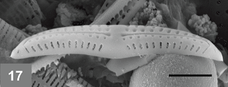
Fig. 3. Microscope images of Medlinella amphoroidea; a) girdle view; b), c), d) valve / face view (Photos: a), c), d) Matt
Ashworth; b) Frankovich et al., 2016; scale bar = 2 μm).
I hope my story will encourage some of you out there to
share your passion for diatoms with other scientists and to pursue any
opportunities that may present themselves. The
seeds of future exciting discoveries start with a conversation. Thanks
again Luca and readers, for our continuing conversations on the
diatom-of-the-month blog.
* Research Faculty at the Southeast Environmental
Research Center, Florida International University.
1. Valvocopulae: the first girdle bands
that attach to the valve
2. Septa: inward projections of silica
that partially separate areas within the cell.
3. Volate pore occlusions: flap-like
outgrowths from the sides of the pores with narrow points of attachment and
irregular branching, as opposed to cribrate or rotate occlusions.


Comments
Post a Comment